|
Wellness – Example projects
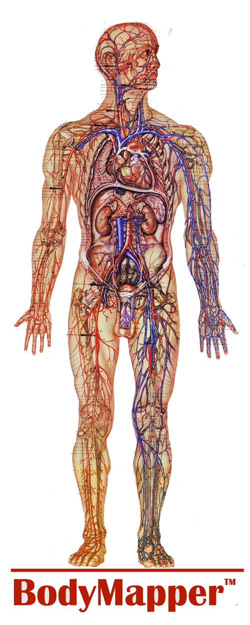
BodyMapping
IRID Inc. conducts basic and applied R&D targeted at specific opportunities to improve the accuracy and cost-effectiveness of medical diagnosis and treatment. Resulting systems and methods address automated identification of patients, standardized personal BodyMapping GIS for Electronic Medical Records, detection and classification of local and systemic conditions from multispectral 2D and 3D imaging. One thrust of the projects is detection of Personal Sentinel Events that may be tipping points in an individual’s physiological or psychological health.
BodyMapping extracts a 3D surface representation of internal anatomical features including cardiovascular and lymphatic elements determined from surface temperature variations. External features, such as wrinkles, moles, sweat pores, and hair follicle, which can be seen in high resolution visible light (VL) images, also can be seen in IR images and form a separate layer of the BodyMap. It is used to precisely align different images of body areas taken over time, in which the temperature, pose, distance, optics, and condition of the subject may be different. The basic novelty of BodyMapping is its use of passive sensor imagery to produce precise high-resolution dynamic maps of subsurface anatomical structures that provide whole-body registration fiducials for automated change detection monitoring.
Compensation for Respiratory Movement
Compensating for respiratory movement during radiosurgery can reduce the area irradiated. Efficacy of irradiation of solid tumors correlates with ability to precisely target the cancer and avoid damaging surrounding healthy tissue. There is especially a need to compensate for breathing movements to better target thoracic and abdominal cancers. 3DIR offers a risk-free method for tracking specific subsurface reference locations during respiration cycles and episodic movements.
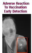 BodyMapper provides consistent wholebody reference mesh over time. This facilitates detection and documentation of changes at specific locations. Applications may include routine screening for early stage pressure ulcers, detecting adverse reaction to vaccination, and monitoring all moles for early detection of changes that may indicate possible melanoma.
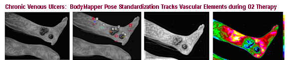
Many research projects consider thermal changes as indicators of wound condition. Use of BodyMapping uniquely provides a method to automatically and precisely register imagery frame-to-frame in order to track microthermal changes along vascular segments in a non-contact and risk-free manner. This technique was used to image chronic non-healing venous ulcers during O2 administration. Evidence of circulation within the wound boundaries could be pinpointed.
 Recent Programs funded by IR&D and DOD, or pending with NIH include:
- Detecting recent use of a psychoactive substance from 3D IR imagery
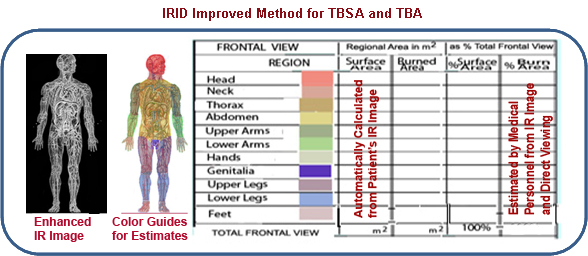
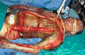
- Burn depth and percent of burned surface determination from 3D IR imaging
- Monitoring wound progression using BodyMapping
- Self-Directed monitoring of moles at walk-in clinics and wellness centers
- Improved Method for calculating Total Body Surface Area and % Burned